Table of Contents
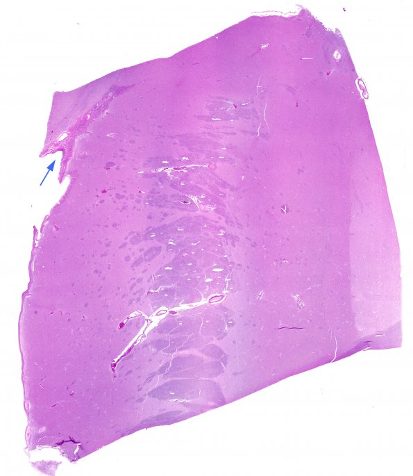
Washington University Experience | BASIC PROCESSES | Ependymal Fusion Seam | 7A1 Ependymal seam, basal ganglia (Case 9) N2 H&E WM
A similar process appears in the lateral ventricles superior to the basal ganglia. In this image the ependymal lining proceedes from the ventricle (arrow) and is fragmented into small tubules and individual cells as it is prolonged into the closure of the ventricle at the superior margin of the basal ganglia (H&E)

