Table of Contents
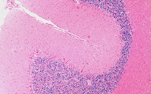
Washington University Experience | BASIC PROCESSES | Neuronophagia | 3A Paraneoplastic cerebellar degeneration (Case 3) H&E 1
3A-G Paraneoplastic cerebellar degeneration. 3A A low magnification image shows areas of apparent Purkinje cell loss, areas of active degeneration of Purkinje cells and hypercellularity of the molecular layer due to increased numbers of microglia, seen at higher magnifications in the next two images (H&E).

