Table of Contents
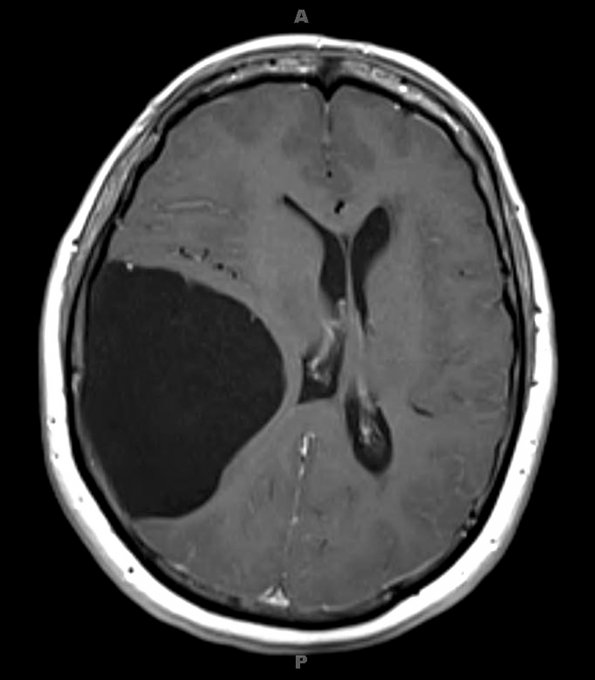
Washington University Experience | DEVELOPMENTAL MALFORMATIONS | Cyst - Arachnoid | 1A1 Cyst, Arachnoid (Case 1) T1 3
1A1-5 The MRI appearance of arachnoid cysts are distinctive with typical broad origins extending from the surface of the brain, although some may appear parenchymal. ---- The T-1 magnetic resonance axial (#1A1), coronal (#1A2) and sagittal (#1A3) images of this arachnoid cyst are isointense with respect to the ventricular CSF. The cyst has compressed and displaced the ipsilateral hemisphere and lateral ventricle.

