Table of Contents
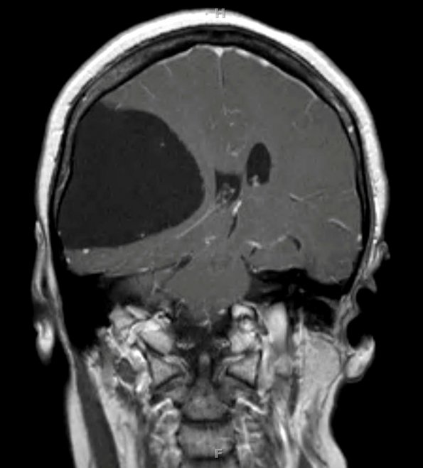
Washington University Experience | DEVELOPMENTAL MALFORMATIONS | Cyst - Arachnoid | 1A2 Cyst, Arachnoid (Case 1) T1 2
The coronal MRI T-1 magnetic resonance image is isointense with respect to the ventricular CSF. The cyst has compressed and displaced the ipsilateral hemisphere and lateral ventricle.

