Table of Contents
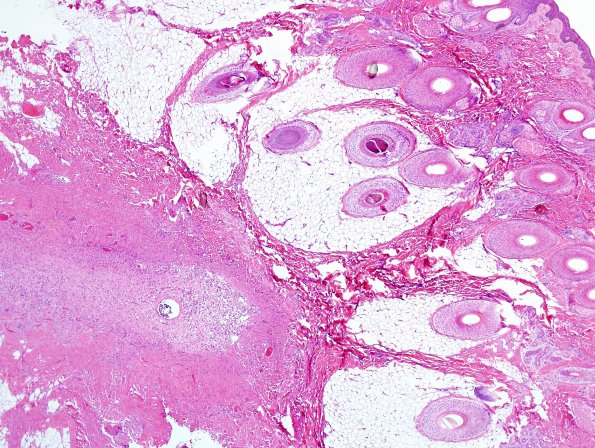
Washington University Experience | DEVELOPMENTAL MALFORMATIONS | Encephalocele | 11A1-Encephalocele-Case-11-H-E-3.jpg
Case 11 History ---- The patient is a five-month-old female who presented with a palpable mass within the midline parietal region. MRI/MRV confirmed an abnormal fetal straight sinus terminating in the region of the midline with an overlying cephalocele. Operative procedure: Excision of occipital-parietal cephalocele. ---- 11A1-3 H&E stained slides of the "occipital-parietal cephalocele" show unremarkable squamous epithelium. There is an ill-defined collection of cells within the deep dermis showing small, round nuclei and abundant foamy pink cytoplasm characteristic of histiocytes intermixed with clusters and whorls of cells with more oval nuclei, reminiscent of meningothelial cells.(H&E)

