Table of Contents
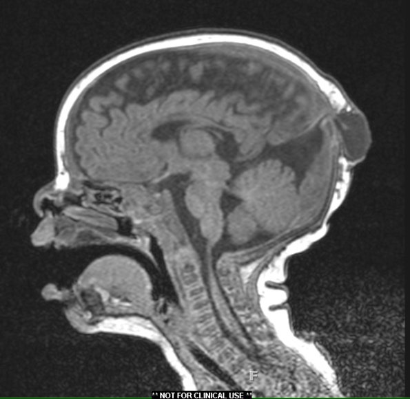
Washington University Experience | DEVELOPMENTAL MALFORMATIONS | Encephalocele | 18A Encephalocele (Case 18) T1 without contrast - Copy
Case 18 History---- The patient is a 3-month-old male who presented with a midline skull lesion. Operative procedure: Excision parietal cephalocele.---- 18A MRI showed a 2.7 cm fluid-filled structure at the midline near the posterior fontanelle, which communicates intracranially along an associated fibrous-appearing stalk with no evidence of herniation of parenchymal tissue into the cystic structure. A midline encephalocele is seen in this T1-weighted image without contrast.

