Table of Contents
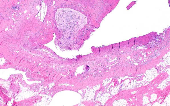
Washington University Experience | DEVELOPMENTAL MALFORMATIONS | Encephalocele | 18B Encephalocele (Case 18) Glial island H&E 1
Routine H&E-stained sections of the "parietal cephalocele" show skin and soft tissue with a central lesion consisting of atrophic epidermis with no adnexal structure and underlying soft tissue containing abundant myxoid stroma. Scattered dilated vascular channels are seen. A small fragment of neuroglial tissue is also present. (H&E) ----Comment: Similar appearing lesions have been variously called “sequestrated meningoceles of the scalp” or “hamartoma of the scalp with ectopic meningothelial elements”. However, due to the intracranial malformation adjacent to the lesion, which is not seen in the two aforementioned entities, as well as the presence of heterotopic neuroglial tissue, we favor this to represent a dysraphic malformative process, most consistent with an encephalocele.

