Table of Contents
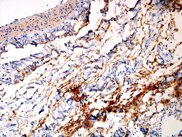
Washington University Experience | DEVELOPMENTAL MALFORMATIONS | Encephalocele | 22E Encephalocoele & (Case 22 EMA
Peripheral to the cystic lining are cleft-like spaces lined by a flattened single cell layer and scattered collections of polygonal cells, suggestive of a leptomeningeal layer. Immunostains were performed and an epithelial membrane antigen (EMA) stain highlights the meningothelial layer. ---- Comment: The histomorphologic and immunophenotypic features are consistent with the diagnosis of meningoencephalocele with immature elements. The latter is an unusual finding in such specimens, but is thought to represent neuroblast-like developmentally appropriate cells reflecting this patient's young age.

