Table of Contents
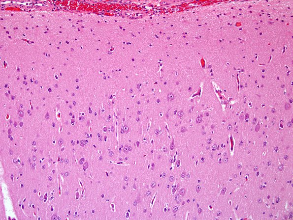
Washington University Experience | DEVELOPMENTAL MALFORMATIONS | Hemimegalencephaly | 1B Hemimegalencephaly (Case 1) A4 H&E
Examination of the cortical resection specimen shows population of all cortical laminae by enlarged, variably dysmorphic neurons which form clumps and rudimentary cortical neuronal columns. Balloon cells are not part of this cortical dysplasia, nor is there evidence of significant polymicrogyria or subcortical heterotopias in this biopsy specimen. Considering the marked increase in cortical size, thickness as well as surface area, and the numerous enlarged neurons, they may constitute the majority of hypertrophic elements in this expanded cortex. (H&E)

