Table of Contents
Washington University Experience | DEVELOPMENTAL MALFORMATIONS | Holoprosencephaly | Alobar HPE | 1A1 Holoprosencephaly (Case 1) 2A
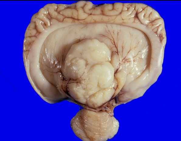
1A1-4 Classic appearance of alobar holoprosencephaly in a 38 week EGA infant who survived only 12 hours after birth. ---- Dorsal view shows a single horseshoe-shaped cerebral hemisphere and ventricle with no evidence of major sulci or lobe formation. The deep gray basal ganglia and thalamus are fused in the midline into a dysplastic lumpy mass. The cerebellum is abnormal in size and structure with expansion of the midline portion.

