Table of Contents
Washington University Experience | DEVELOPMENTAL MALFORMATIONS | Hydranencephaly | 3E5 Hydranencephaly (Case 3) N14 H&E A2
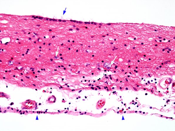
This image is the typical appearance of the membranous residua. The subarachnoid space (arrowheads), the ventricle/ependymal surface (blue arrow marks cilia) and residual glial tissue enclosing scattered calcifications (white arrows). Considering the enormous membranous surface from the cortex converted in response to the tissue loss, there has been a proliferation of ependyma to cover much of the membranes.

