Table of Contents
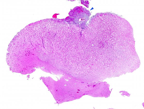
Washington University Experience | DEVELOPMENTAL MALFORMATIONS | Meningioangiomatosis | 7B1 Meningioangiomatosis (Case 7) H&E WM copy
7B1-5 Hematoxylin and eosin stained sections show fragments of cortex with fine trabeculae of perivascular meningothelial cells extending from the cortical surface into the brain parenchyma up to the white matter junction. The cells are overall bland. No mitoses are appreciated. The intervening parenchyma shows some reactive changes. Adjacent neurons are large and reactive with mild atypia. At the cortical surface there is an associated small meningothelial cell proliferation (arrowhead, 7B1) but this is not brain invasion by a meningioma.

