Table of Contents
Washington University Experience | DEVELOPMENTAL MALFORMATIONS | Polymicrogyria | 1B1 Polymicrogyria (Case 1) H&E whole mount 2
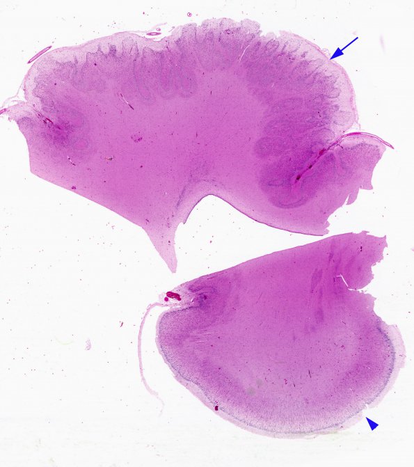
These two cortical whole mount images of the case shown grossly in #1A previously allow the comparison of the polymicrogyric cortex (arrow) and the more normal cortex (arrowhead) in adjacent areas. Notice that the polymicrogyric area is typically thicker than normal cortex. True pachygyric areas are rarer than simple thickened cortex and more likely represent polymicrogyria. (H&E)

