Table of Contents
Washington University Experience | EPILEPSY | Hemiatrophy | 1 Hemiatrophy with Seizures (Case 1)
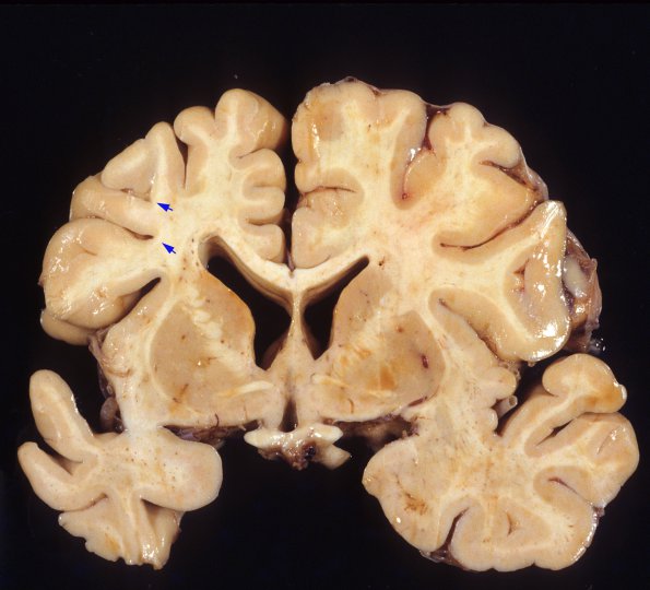
In this elderly patient with poorly characterized chronic seizures, the left ventricle is dilated and blunted. The white matter adjacent to the left ventricle shows discoloration and granularity. There also appears to be local loss of cortical gray matter at the depths of the sulci (arrows), a histologic picture of ulegyria. It is likely that there is a superimposition of hypoxia/ischemia also in this case, and makes it less likely to represent a pure process.

