Table of Contents
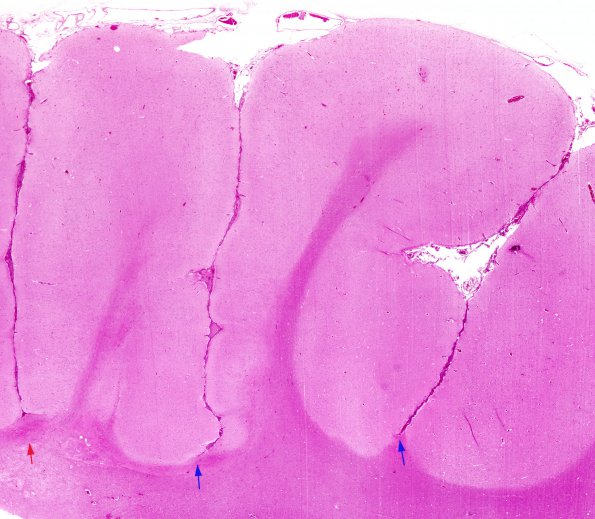
Washington University Experience | EPILEPSY | Ulegyria | 1B2 Ulegyria (Case 1) H&E whole mount 2
The red arrow at the left margin shows the area reproduced at higher magnification in the following images in which it is stained for myelin and axons. Other foci of ulegyria are designated with blue arrows (H&E).

