Table of Contents
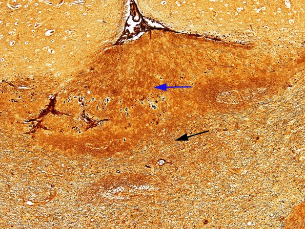
Washington University Experience | EPILEPSY | Ulegyria | 1C2 Ulegyria (Case 1) Biels 2
Axons in the depths of this sulcus are markedly diminished in number (blue arrow) in comparison with adjacent white matter with axons (black arrow). Higher magnification (in 1C3) confirms the lack of axons. Comparison of these images with the #1C1 image shows that the tissue which stains for myelin contains no axons. It has been reported than hypoxic/ischemic lesions in neonates may exhibit myelination of glial processes rather than axons (Bielschowsky).

