Table of Contents
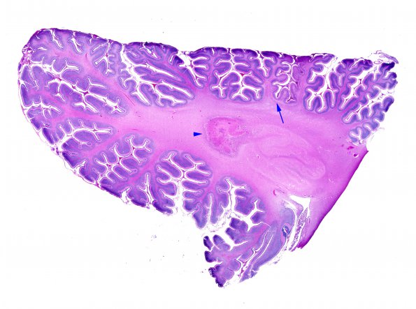
Washington University Experience | INFECTION | Bacteria | Meningitis | Meningitis, complications - acute | 1G1 Meningitis, Group B STR (Case 1) N9 H&E whole mount copy
There is prominent involvement of the cerebellum in two patterns: ---- The first pattern consists of granule and Purkinje cell loss which is enhanced in the depths of the sulci (arrow, 1G1) in particular. Higher magnification of the region of the arrow in image #1G1 (in 1G2,3) shows marked loss of Purkinje and granule neurons with Bergmann astrocytosis, and endothelial prominence. A higher magnification demonstrates a prominent mineralized Purkinje cell (arrow, 1G3). Loss of neurons in the depths of sulci is a typical pattern of hypoxia/ischemia.

