Table of Contents
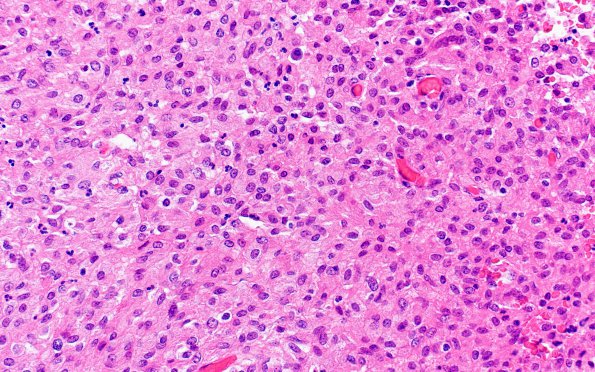
Washington University Experience | INFECTION | Bacteria | Mycobacteria, Atypical | 2A5 Mycobacterium, atypical MAI (AANP1998, Case 3) H&E 4
This region of the field in image 2A3 shows numerous epithelioid cells with granular cytoplasm, prominent cell borders and no hints of neoplasia or mitotic activity (H&E). (AANP Diagnostic Slide Session 1998, Case 3)

