Table of Contents
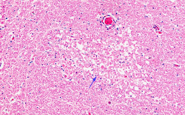
Washington University Experience | INFECTION | Bacteria | RMSF | 2D1 RMSF (Case 2) H&E 40X 6 copy
2D1,2 Both perivascular and parenchymal collections of axonal spheroids (arrow, 2D1) and microglia complete the microscopic image

