Table of Contents
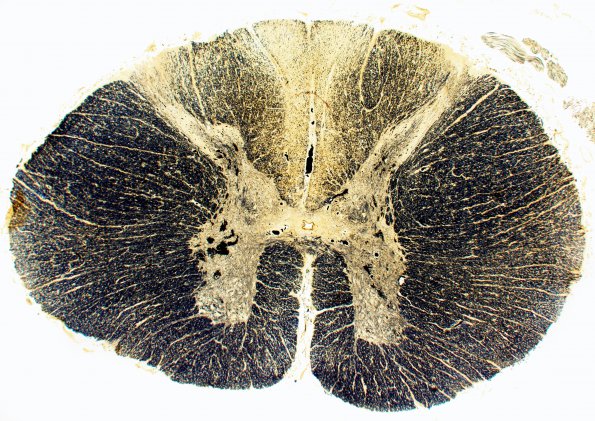
Washington University Experience | INFECTION | Bacteria | Syphilis | 4 Tabes dorsalis, spinal cord (Case 4) Weil's myelin stain 1
This image shows a section of the spinal cord stained with Weil's stain demonstrating loss of individual myelinated axons in the dorsal columns of a patient with tabes dorsalis. The gracile portion of the dorsal columns is more substantially depopulated than the adjacent cuneate fasciculus. Each preparation shows significant loss of myelin in the dorsal columns, reflecting the loss of DRG neurons and/or damage to ascending large fibers.

