Table of Contents
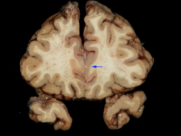
Washington University Experience | INFECTION | Bacteria | TB - Individual Case | 1F9 TB Meningitis, Ventriculitis, Tuberculoma, Rich foci (Case 1) Gross_33 copy
A single intracortical tuberculoma (arrows, 1F9,10) extends to the subarachnoid space, which is better seen in #1F10 (arrow). This pattern has been called a “Rich focus” and is thought to represent the point of initiation of tuberculous meningitis after hematogenous spread to the parenchyma.

