Table of Contents
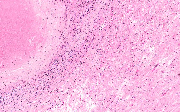
Washington University Experience | INFECTION | Bacteria | TB - Meningitis & Tuberculoma | 4B3 Abscess (Case 4) 4X H&E 2
The lesion appears to be a forming a classical abscess with a central necrotic area on the right portion of the image, surrounded by granulation tissue/inflammation, reactive brain parenchyma and a paucity of multinucleated giant cells. This image shows the trilaminar constituents of the abscess.

