Table of Contents
Washington University Experience | INFECTION | Fungus | Blastomycosis | 2A3 Blastomycosis (Case 2) H&E 13 copy
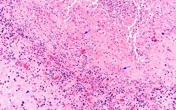
This granuloma shows necrosis in the upper right corner, epithelioid histiocytes and multinucleated giant cells (arrows), spindled fibroblasts and granulation tissue, frequent lymphocytes and plasma cells, and a developing capsule (arrowhead) in the left corner (H&E).

