Table of Contents
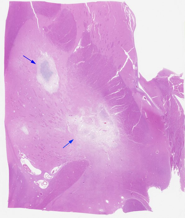
Washington University Experience | INFECTION | Protozoa | Toxoplasmosis | 1C1 Toxoplasmosis Rx (Case 1) H&E whole mount 2
1C1-3 Basal ganglia pathology (arrows) consist of necrotizing lesions characterized by lymphocytes, macrophages, mononuclear and polymorphonuclear inflammatory cells, reactive astrocytes, and microglia with no significant capsule formation. The central focus of the superior lesion, #1C2) consists of dystrophic calcifications of various sizes which accumulate in macrophages (arrows, #1C3) where they may mimic (poorly) the appearance of toxoplasma cysts (H&E).

