Table of Contents
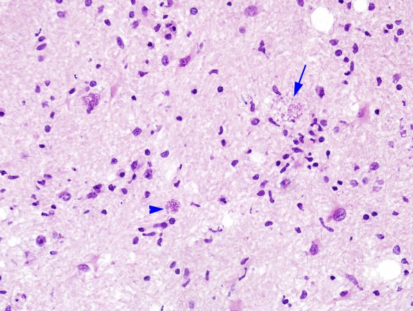
Washington University Experience | INFECTION | Protozoa | Toxoplasmosis | 3A1 Toxoplasmosis (Case 3) H&E 3A
3A1-4 In this case individual lesions are more easily identified by H&E, ranging from bradyzoites in cysts (arrowhead) and tachyzoites liberated from cysts (arrow) seen at higher magnification in #3A2 and #3A3 (H&E). The substructure of tachyzoites (arrow, 3A4) within the parenchyma is shown.

