Table of Contents
Washington University Experience | INFECTION | Viruses | CMV | CMV, Congenital-Neonatal | 1A1 CMV (Case 1) 1 Intrauterine vs normal ExpA
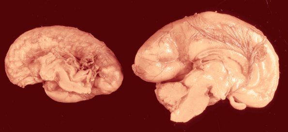
This image is the comparison of the markedly atrophic fetal brain infected with CMV on the left with a normal 24 week gestation brain on the right.

