Table of Contents
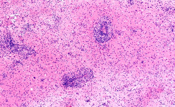
Washington University Experience | INFECTION | Viruses | PML (JC Virus) | PML - Enigmatic Case | 1B1 PML (Case 1) H&E 10X 1
1B1,2 Hematoxylin and eosin stained permanent sections of the neurosurgical specimen show brain parenchyma with increased cellularity. There is increased parenchymal and perivascular mixed inflammatory infiltrate composed of lymphocytes, histiocytes, and plasma cells. Mitoses are not frequent. There is no microvascular proliferation or pseudopalisading necrosis. The histopathology was remarkable for substantial perivascular inflammatory cells suggesting a wide differential diagnosis including vasculitis (possibly collagen vascular), lymphoma, inflammatory demyelination, and, particularly with the atypical appearance of astrocytic elements, glioma.

