Table of Contents
Washington University Experience | INFECTION | Viruses | PML (JC Virus) | PML - Gross Pathology | 10A3 PML (Case 11)_12
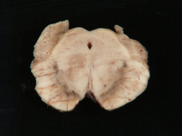
10A3 A discrete brainstem lesion is shown in gross appearance (10D) as well as whole mounts of H&E (#10B1), LFB-PAS (#10B2) and a Bielschowsky silver stain for axons (10B3). This lesion showed pallor in H&E and loss of myelin stain in the presence of maintained axons, the characteristic staining of demyelinative lesions. Nonetheless, PML not uncommonly demonstrates a degenerative component and axonal spheroids.

