Table of Contents
Washington University Experience | INFECTION | Viruses | PML (JC Virus) | PML - Illustrative Case | 1H1 N1 PML N6 H&E 100X 4 copy
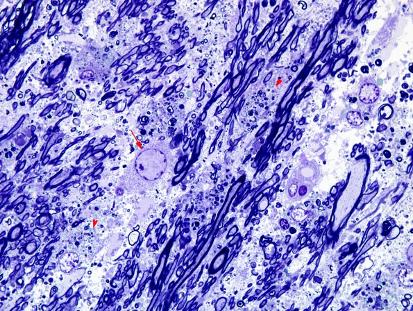
This image shows relatively well preserved myelin sheaths on many of the axons admixed with some myelin debris in macrophages (arrowhead). An enlarged oligodendrocyte (arrow) shows expansion of the nucleus. (One micron thick toluidine blue stained plastic section)

