Table of Contents
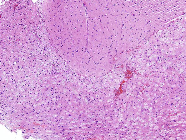
Washington University Experience | INFECTION | Viruses | PML (JC Virus) | PML - Microscopic Pathology | 3A1 PML (Case 3) H&E 5
3A1,2 There is clear separation of gray matter at the superior border of the image from the white matter below with its numerous macrophages and enlarged, oligodendroglial "plum colored" cells, seen at higher magnification in #3A2.

