Table of Contents
Washington University Experience | INFECTION | Viruses | PML (JC Virus) | PML - Microscopic Pathology | 3A2 PML (Case 3) H&E 4
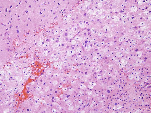
There is clear separation of gray matter at the left superior border of the image from the white matter below with its numerous macrophages and enlarged, oligodendroglial "plum colored" cells.

