Table of Contents
Washington University Experience | INFECTION | Viruses | PML (JC Virus) | PML - Neurosarcoid vs PML Case | 2C PML (Case 1) NF 40X
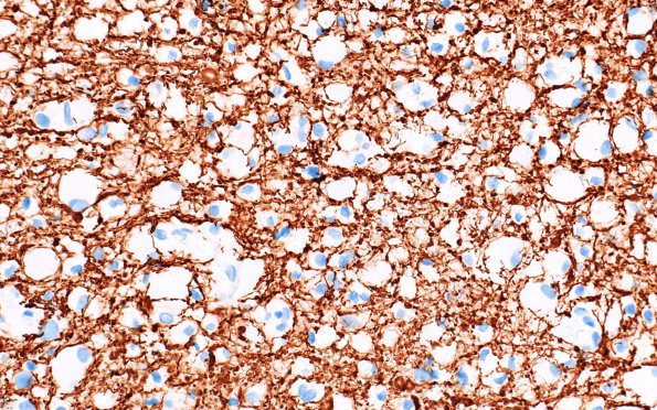
Numerous axonal processes are identified in this neurofilament stain of the area shown previously as image #2B1. Many of the axons are cut in cross section. This area of myelin loss continues to show numerous preserved axons and has very few axonal spheroids, .i.e., degenerative swollen axons containing accumulated cytosomes. (NF IHC)

