Table of Contents
Washington University Experience | INFECTION | Viruses | PML (JC Virus) | PML - Ultrastructure | 1A1 PML (Case 2) Plastic 1a copy
Case 1 History
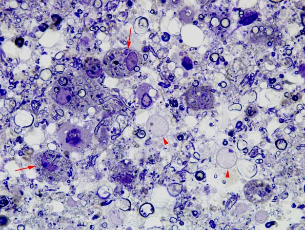
One micron thick toluidine blue stained plastic embedded section of a white matter biopsy shows only scattered normal myelinated axons at a site where large numbers should be demonstrable. There are numerous lipid laden macrophages (arrow) and two oligodendrocytes with large pale PML-infected nuclei (arrowheads) (1 micron thick toluidine blue plastic section)

