Table of Contents
Washington University Experience | INFECTION | Viruses | PML (JC Virus) | PML - Granule Cell Neuronopathy | 1F1 PML, R cerebellum (Case 1 Autopsy specimen) _10
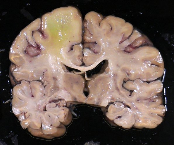
Gross findings at brain autopsy performed on the patient whose neurosurgical biopsy has been illustrated in the previous images. There was no classical PML pathology in any white matter area of the cerebral hemispheres or brainstem. The green discoloration reflects the breakdown of the blood brain barrier in the presence of circulating hyperbilirubinemia due to the metastasis (not determined which site was the origin but lung was most likely) in immediately adjacent brain slice (#1F2). A microscopic image of the tumor is shown in image #1F3 (H&E).

