Table of Contents
Washington University Experience | INFECTION | Viruses | Rubella | 1E Rubella (Case 1) Hx Childhood Adult at death gross 3
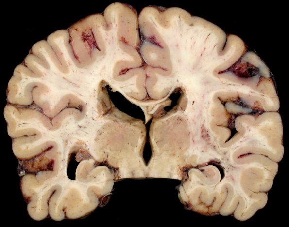
The brain appears atrophic with expansion of several sulci, dilatation and blunting of the lateral ventricles, shrinkage of the temporal lobe with dilatation of the temporal ventricular horns and impressive thinning of the corpus callosum. The optic tracts are hard to visualize. The white matter at the edges of the lateral ventricle shows discoloration and, in some cases, frank tissue loss which may reflect periventricular neonatal hypoxic/ischemic injury. There are scattered strands of tissue bridging part of the lateral ventricles.

