Table of Contents
Washington University Experience | METABOLIC | Kernicterus | 8A4 Kernicterus (Case 8) 3 (2)
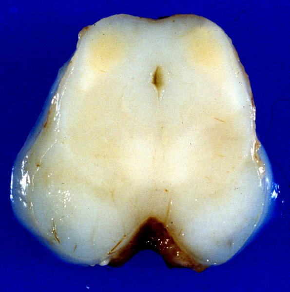
Multiple brain nuclei show yellowish discoloration including basal ganglia, especially globus pallidus, thalamus, and dorsal aspects of the midbrain show prominent yellow discoloration: ---- Additional findings (not shown): Within the subarachnoid space around the cord were many polymorphonuclear leukocytes, monocytes and much necrotic debris. Tissue gram stain revealed Group B streptococci; some phagocytic cells which appeared to be totally filled with bacteria. Multiple small subependymal hemorrhages and small periventricular infarcts were located adjacent to the lateral ventricles consistent with hypoxic/ischemic encephalopathy

