Table of Contents
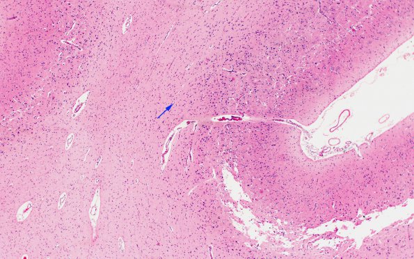
Washington University Experience | METABOLIC | Menkes disease | 1F2 Menke's Dz (Case 1) P Temporal lobe H&E areaA 3 copy
Increased magnification of the designated area of image 1F1 (H&E) shows the separation of the atrophic cortex from the underlying gliotic white matter (arrow) (H&E)

