Table of Contents
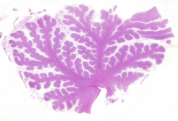
Washington University Experience | METABOLIC | Menkes disease | 1H1 Menke's Dz (Case 1) 1 H&E whole mount 2
1H1-6 These images of the cerebellar vermis are characterized by subtotal granule cell loss, a relatively well maintained number of Purkinje cells and even patchy preservation of the fetal external granule cell layer (arrow, 1H4) in this 14 month old child. The molecular layer appears disordered at this magnification particularly 1H5. (H&E)

