Table of Contents
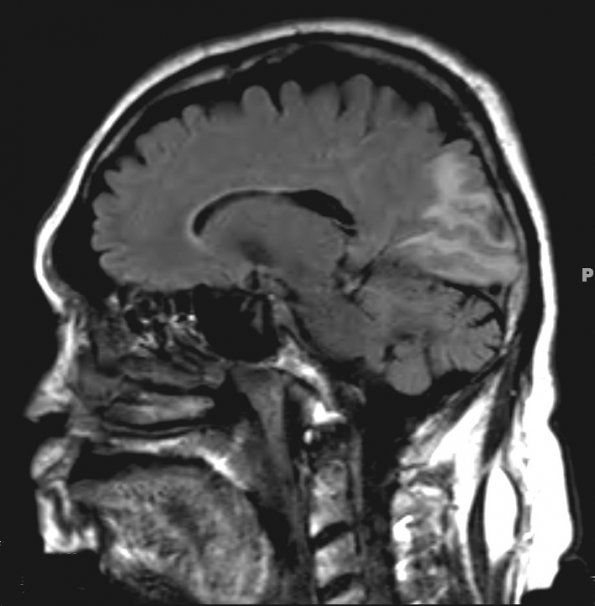
Washington University Experience | MISCELLANEOUS | Amyloidoma | 1A1 Amyloidoma, cerebral (Case 1) FLAIR - Copy
1A1-6 MRI images ---- 1A1,2 FLAIR (1A1) and TIRM BLADE (1A2) MRIs show hyperintensity in the left occipital lobe which appears confined to the white matter without significant mass effect. MRI appearances may vary from case to case.

