Table of Contents
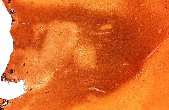
Washington University Experience | MYELIN (IMMUNE-MEDIATED) | ADEM | 1D1 ADEM (Case 1) Bielschowsky A 2X
1D1-4 Although the pale perivascular collars suggest axon loss in this Bielschowsky silver stain, higher magnification images show numerous intact axons and relatively few axonal spheroids, evidence of relative axonal preservation compared to more substantial myelin loss. The density of axons also reflects intervening reactive cell infiltrate. (Bielschowsky silver stain for axons)

