Table of Contents
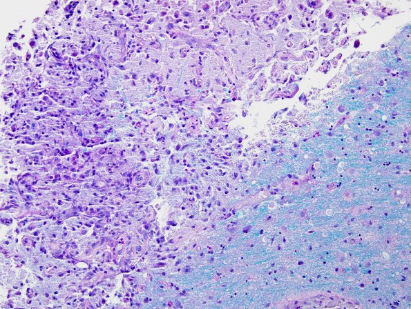
Washington University Experience | MYELIN (IMMUNE-MEDIATED) | ADEM | 1D6 Infarct (Case 6) MS LFB-PAS 40X
1D6,7 For comparison, this is the margin of a subacute infarct showing myelin loss (1D6) and, in the next image (1D7) the same area stained for axons with the Bielschowsky silver stain showing loss of axons in the center (arrowheads) of the lesion and dystrophic axonal swellings at its margins (arrows)

