Table of Contents
Washington University Experience | MYELIN (IMMUNE-MEDIATED) | Balo Concentric Sclerosis | 2C1 Balo Concentric Sclerosis (Case 2) Composite 2
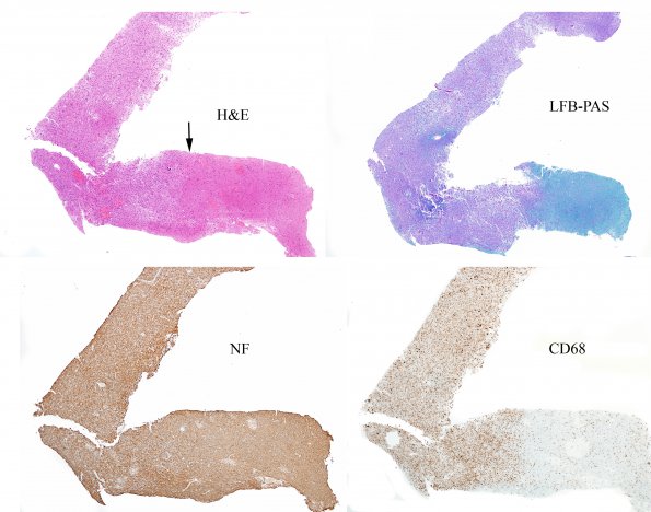
The surgical biopsy consisted of a number of fragments of subcortical white matter of which one was particularly useful. This is a composite image of near sequential sections of tissue stained for H&E, LFB-PAS (myelin), neurofilament IHC (axons) and CD68 (macrophages). The arrow identifies a segment which is the border between the demyelinating process and normal white matter. Additional sections of this area will be presented at higher magnification in the following images.

