Table of Contents
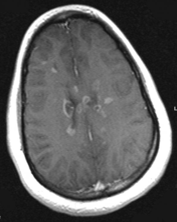
Washington University Experience | MYELIN (IMMUNE-MEDIATED) | MS - Cerebral Cortex | 2A1 MS (Active Lesions) (Case 2) T1 Post-Contrast - Copy
2A1-3 MRI examination ---- 2A1,2 These T1-weighted images with contrast show numerous plaques in the periventricular region as well as partial contrast enhancing lesions which appear to involve the depths of sulci (arrows, 2A2)

