Table of Contents
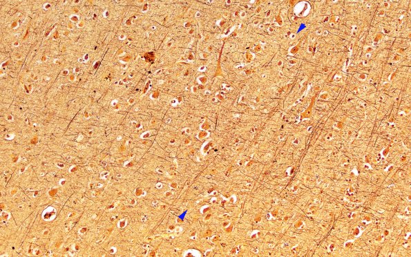
Washington University Experience | MYELIN (IMMUNE-MEDIATED) | MS - Cerebral Cortex | 2H MS Cortical Plaque (Case 2) from PLP junction area Biels 20X 1 copy
The margin (between arrowheads) of demyelinated (left portion of the image) and normally myelinated (right side of image) cortex shows slight decrease in axons in this Bielschowsky stained preparation.

