Table of Contents
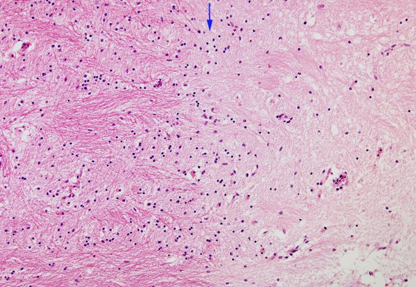
Washington University Experience | MYELIN (IMMUNE-MEDIATED) | MS - Cerebral Hemispheres | 1B4 MS, Plaque (Case 1) H&E 20X copy
1B4-6 Higher magnification of the wall of the plaque stained routinely (H&E, 1B4), for myelin (1B5, LFB-PAS) and axons (1B6, Bielschowsky). ---- The plaque edge (arrow) is shown. The plaque itself is pale and hypocellular with few residual oligodendroglia, minimal inflammatory cells and only rare macrophages, i.e., it is largely inactive. The majority of the residual cells within the plaque are astrocytes and endothelial cells of the microvasculature.

