Table of Contents
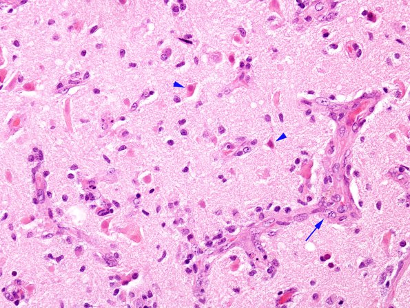
Washington University Experience | MYELIN (IMMUNE-MEDIATED) | MS - Cerebral Hemispheres | 1C1 Infarct (Case 1A) compare with MS H&E 40X copy
1C1-3 In some settings plaques may resemble ischemic lesions. In this series of images the edge of a subacute infarct is examined with routine stains (H&E, 1C1), for myelin (LFB-PAS, 1C2) and axons (Bielschowsky, 1C3) ---- The margin of this infarct which involves both white and gray matter shows eosinophilic neuronal necrosis (arrowheads) and endothelial prominence in the microvasculature (arrow) (H&E)

