Table of Contents
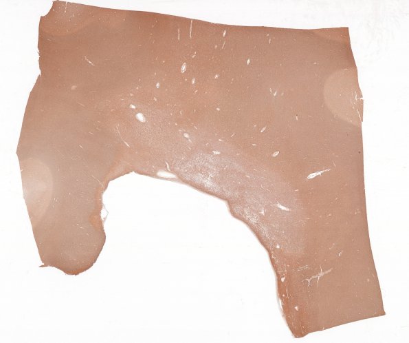
Washington University Experience | MYELIN (IMMUNE-MEDIATED) | MS - Cerebral Hemispheres | 7B5 MS Plaque (Case 7) whole mount plaque Correlate with N8 NF 1
7B5-8 These images compare the two stains used for axons. Neurofilament IHC (7B5) demonstrates a well preserved axonal complement, seen at higher magnification in 7B6 (NF IHC). In comparison, the Bielschowsky stain (7B7) is more pallid in the demyelinated sector, suggesting more substantial axon loss. In reality, the Bielschowsky stain results in increased non-axonal background in intact white matter. Higher magnification demonstrates numerous residual axons in the demyelinated focus (#7B8).

