Table of Contents
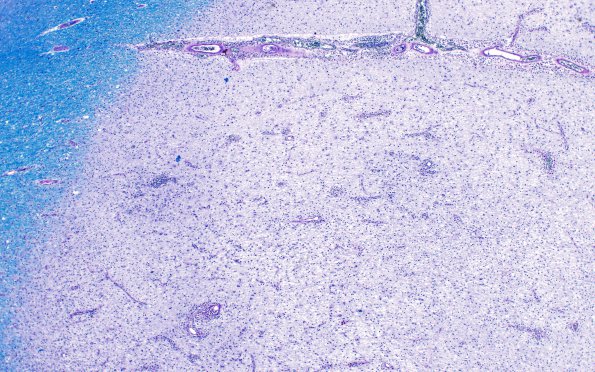
Washington University Experience | MYELIN (IMMUNE-MEDIATED) | MS - Chronic Active Plaque | 2E2 MS, chronic active (Case 2) N1A Edge LFB-PAS 4X
2E2-7 This series of images of the active demyelinating area at different magnifications shows the loss of myelin. The border with the normal white matter is a bit more diffuse than in an inactive lesion. There are astrocytes, lymphocytes, residual oligodendrocytes, macrophages containing either blue particles (early phagocytosis of myelin) or red granules (metabolized myelin, best in 2D7) which surrounds the vasculature in the Virchow-Robin spaces (LFB-PAS)

