Table of Contents
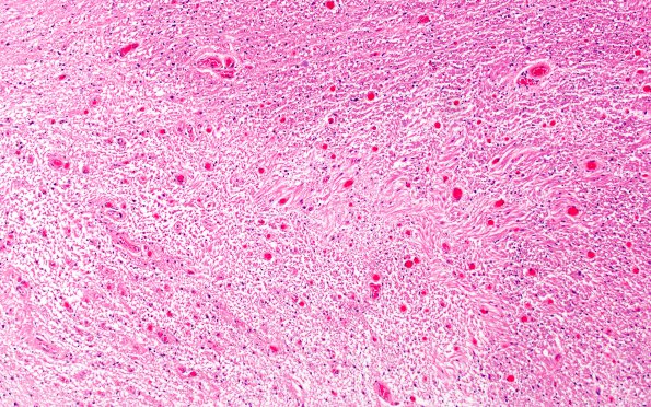
Washington University Experience | MYELIN (IMMUNE-MEDIATED) | MS - Optic Nerves | 1F1 MS, optic nerve (Case 1) Area C H&E 10X
1F1-3 The area identified with an arrow in image 1B1, representing the junction of the demyelinated plaque with the adjacent myelinated chiasm, is shown stained with H&E (#1F1), for myelin (#1F2, LFB-PAS) and axons (#1F3, NF IHC)

