Table of Contents
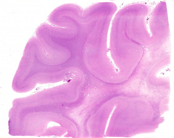
Washington University Experience | MYELIN (IMMUNE-MEDIATED) | MS - Primary Progressive (PPMS) | 1D1 MS, Primary Progressive (PPMS) (Case 1) H&E N12 WM
1D1-10 The following series of cortical images were derived from the right occipital lobe. ---- 1D1-3 This series of images includes tissue stained by H&E (1D1), LFB-PAS for myelin (1D2), and Bielschowsky for axons (1D3). The subcortical white matter, in particular, appears to be grossly depopulated of axons.

